
Contact Info

Interventional Radiology

About Interventional Radiology
 

Interventional Radiology is a branch of medicine which is devoted to treating various vascular (other than the heart) and non-vascular conditions, through integration of clinical and imaging based diagnosis and minimally invasive therapy. These procedures are performed by trained Interventional Radiologists.

Interventional radiologists are doctors who with the help of mainly specialized X-ray machines, use their expertise to guide tiny instruments, such as catheters, through blood vessels or through the skin to treat various diseases without surgery.

Interventional radiologists are specialists who have trained rigorously in minimally invasive interventions using imaging guidance; they combine their imaging background with clinical knowledge to treat various vascular and non-vascular pathologies.

With their expertise in imaging and the specialized and advanced training only in the field of non-surgical techniques with imaging guidance, interventional radiologists can treat a large number of ailments within the body by delivering the treatment directly to the source of the problem.

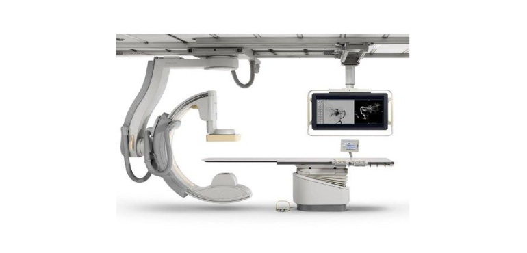
Breach Candy Hospital has a state of the art Interventional radiology suite. The Philips Allura Clarity FD 10 has high resolution digital subtraction images, with low radiation. Adjunctive machines such as peripheral laser, intravascular ultrasound, doppler are part of the department and greatly enhance the procedural results.

With the availability of the best imaging modalities (USG, CT, MRI, PET scan) and highlyt trained Interventional Radiologists (IR’s), the interventional radiology unit of Breach candy is one the best in the country.

Read More

Services

  • Thrombectomy for Acute Stroke.
  • Carotid stenting, Intracranial stenting.
  • Intracranial aneurysms Coiling, Flow Diversion.
  • Embolization of brain AVM, AVF, Dural A-V Fistula.
  • Spinal Cord Vascular Malformation (AVM, AVF)
  • Vertebroplasty
  • Maxillofacial Vascular Malformation
  • Paediatric Neurointerventions (Vein of Galen Aneurysmal Malformation, Pial AVF and Dural Sinus Malformation).

PERIPHERAL VASCULAR DISEASE:

Peripheral vascular disease is due to plaques causing blockages in the arteries supplying the lower limbs (legs). This can lead to pain on walking (claudication), pain during lying down (rest pain) and non healing ulcers and gangrene of the feet. Interventional radiology plays an important role in treating these blockages (stenosis/occlusion), by angioplasty/stenting. Various state-of-the-art techniques such as balloon angioplasty, stenting, stent grafts, drug eluting ballooning and stenting, thrombolysis, laser, shockwave lithoplasty are used to clear these blockages and relieve the symptoms.

AORTIC ANEURYSMS AND DISSECTIONS :

When an artery increases in size it can burst and cause life threatening bleeding, this condition is known as an aneurysm of the blood vessel. When this occurs in the main blood vessel, the aorta, it is known as aortic aneurysms. When the aorta, undergoes an internal tear, it is known as an aortic dissection.

Earlier these required surgical treatment which was associated with a moderate risk. Endovascular aortic stent grafting is now one of the main modalities for treating aortic aneurysms and dissections.

Here, stent grafts are used, which are stents with a surgical graft sutured on them. These devices exclude the aneurysm from the circualation, which prevents them from rupturing. Standard thoracic and abdominal aortic aneurysms, thoracoabdominal aortic aneurysms, arch aneurysms, dissections can now be treated with standard endovascular stent grafts, fenestrated aortic stent grafts. The advantages of these procedures are no or shortened anaesthesia time, quicker recovery and lower risk.

VARICOSE VEINS :

Varicose veins are twisted, enlarged veins of the superficial venous system, mainly of the legs. This common disorder can cause cosmetic concern, hyperpigmentation of the legs, non healing venous ulcers. The treatment for these can be medical (compression stockings, exercise) or endovenous; where we use laser, radiofrequency ablation or venaseal vein glue technique to close the varicosities and achieve ulcer healing. This treatment is highly effective and allows the patient to get back to normal activity within a day.

DEEP VEIN THROMBOSIS :

Clots can form anywhere in the vascular system, with the most common location being the deep veins of the lower limbs, which is known as deep vein thrombosis. These clots (thrombii) lead to venous swelling, ulcers and can lead to life threatning pulmonary thromboembolism. The treatment earlier was only medical, by giving anticoagulants, which was not satisfactory for large thrombii. Endovascular modalities such as IVC filters, suction thrombectomy (Penumbra, Angioget), thrombolysis, venous stenting for chronic deep venous thrombosis and May Thurner syndrome, play an important role in removing the thrombi from the deep veins of the legs and pulmonary arteries, hence relieving limb swelling, breathlessness.

INTERVENTIONAL ONCOLOGY :

Certain type of cancers can be treated by interventional radiology techniques. Most common are types of liver cancers, such as, hepatocellular carcinomas (HCC’s), colorectal liver metastasis, bile duct carcinoma (cholangiocarcinomas). These are treated by specialized techniques- tranarterial chemoembolization (TACE) , transarterial radioembolization (TARE), radiofrequency/microwave/cryoablation. These procedures are used for tumour control for inoperable tumours, enhance the quality of life for inoperable lesions, reduction in size of the tumour to facilitate surgery/cure.

Transarterial chemoembolization (TACE) is a procedure in which a low dose chemotherapy agent if delivered via the arterial route, targeted into the liver tumour, followed by reduction in the blood supply to the tumour by particulate embolization. Transarterial Radioembolisation (TARE) utilizes delivery of yttrium 90 (Beta radiating agent) directly into the tumour bed, via the arterial route to irradiate the tumour.

Ablative techniques such as radiofrequency, microwave and cryoablation are used to either burn (radiofrequency, microwave) or freeze (cryo) the tumour, so as to kill the tumour cells and try and achieve tumour control.

Procedures such as bile duct drainage and stenting for inoperable bile duct tumours, DJ stenting for inoperable urinary tract tumours, radiofrequency ablation for inoperable lung and kidney tumours are also done.

EMBOLISATION PROCEDURES :

Certain conditions such as uterine fibroids, benign prostatic hypertrophy, varicoceles, arterial and venous malformations can be treated very effectively with an interventional technique known as embolization. Here the blood supply to these lesions are blocked superselectively, to reduce the size of the lesion, allievate symptoms caused by these conditions. The effectiveness and results of these procedures are excellent and well proven.

Advantages of Interventional Radiology :

Although no medical treatment is 100% risk free, the risks of these interventional radiological procedures are far lower than the conventional form of treatment which used to be open surgery.

All of these procedures are performed with just a small puncture in the vessel, hence obviating the need of large incisions.

Most of these procedures require only 1-2 days of hospital stay as compared to surgical procedures which require 7-8 days.

Majority of the procedures are performed under local anesthesia as compared to open surgery which requires general anesthesia.

Risk, pain and recovery time are often significantly reduced.